


By Brinter Ltd.
Brinter® Tissue Models for Cancer Research
Brinter® offers an accessible gateway into the exciting new world of 3D bioprinting, the revolutionary technology that has exciting applications in cancer research and therapies.
Bioprinting, based on extrusion-based printing but using biomaterials (bioinks), allows researchers to fabricate geometrically well-defined 3D scaffolds seeded with cells rapidly, inexpensively and reproducibly. This technology can meet the urgent need for better and more affordable pre-clinical human tissue models that more accurately represent human cancer pathways in realistic biological contexts.
Bioprinting in-vitro tumor models based on human cancer cells can accurately reproduce the characteristics of human cancer tissue, allowing faster studies in complex interactions such as cancer cell dynamics during vascularization or metastasis of cancer cells. Bioprinted tumor models that use a patient’s own cancerous cells can also enable fully personalized anticancer drugs.
Tissue Models for Cancer Research
Cancer is a leading cause of death worldwide in countries of all income levels. Furthermore, the number of cancer cases and deaths is expected to grow rapidly as populations grow, age, and adopt lifestyle behaviors that increase cancer risk. It is estimated that more than 20 million new cancer cases annually will occur by 2025.
Cancer therapeutics currently have the lowest clinical trial success rate of all major diseases. The majority of cancer research is conducted in mouse models that do not accurately represent human tumor biology. Furthermore, two-dimensional tumor models lack cell-cell and cell-matrix interactions present in 3D. They do not precisely represent a physiologically relevant environment.
Cancer research applications
Better and more affordable pre-clinical human tissue models are urgently needed to representing more accurately human cancer pathways in realistic biological contexts.
Bioprinting can recreate complex cancer microenvironments by precisely situating multiple cell types and even microcapillaries. This approach will benefit the study of cancer pathogenesis and metastasis.
In the future, bioprinted tumor models created using a patient’s own cancerous cells could also enable the personalization of anticancer drugs.
Bioprinting techniques in cancer research
Brinter® 3D bioprinting platforms use revolutionary 3D biomanufacturing technology to drive the field of 3D cell culture models, regenerative medicine and tissue engineering into the future.
Brinter® bioprinter setups can be individually configured to specific research and manufacturing applications ranging from personalized tissue models for drug screening to printing complex drug formulations and ultimately to bioprinted entire functional organs from patients’ own cells for those in need of organ transplant.
Specific techniques include:
- Creating 3D tumor models for cancer research: Brinter’s automated fabrication process allows a researcher to create complex 3D tumors in vitro by printing reproducible cancer models by carefully placing various cell types and supporting biomaterials to construct physiologically relevant cancer models across well plates in a high-throughput manner. These models allow studies concerning complex interactions such as cancer cell dynamics during vascularization or metastasis of cancer cells.
- Fully personalized models: Bioprinted tumor models can even be created using a patient’s own cancerous cells, enabling full personalization of anticancer drugs, along with Drug Sensitivity and Resistance Testing (DSRT). Brinter’s brand-new integrated slicer feature allows full control of the various printouts during a single print job by setting control and experimental groups on the well plates or Petri dishes with just a few button clicks, allowing multiple anticancer drugs to be tested simultaneously.
- Full reproducibility: Brinter® automated precision printing technologies allow cancer models to be effortlessly printed, multiplied and repeated on any cell culture platform. Research quality improves due to reduced human error, as the production is done automatically in a controlled environment.
Matching Bioprinter to cancer applications
Brinter recognizes that 3D bioprinting is a fast-evolving and revolutionary technology in which each new study and application needs to feed back into design and in which each individual bioprinter platform needs to be configurable to specific user needs.
This has inspired a highly modular approach to Brinter® design and fabrication in which each Brinter® Bioprinter is effectively custom-built and infinitely adaptable with a wide choice of individual tools and features.
The upcoming Brinter® technology features sets of proprietary fluidic controllers, actuators, high-frequency solenoids and valves, with precision sensors to control multiple flow parameters and manipulate multiple fluids individually for active mixing, flow focusing, sorting, and structure generation.
This creates a highly adaptable bioprinter platform that can be used as a versatile tool for basic research across a variety of oncology applications.
Cancer research Brinter® technologies
Depending on the specific application, different combinations of Brinter® technologies can be used, such as:
- Pneuma Cooled print head: Pneumatic print head with cooling for printing low to medium viscosity hydrogels, silicones and pastes, such as collagen or Matrigel.
- Pneuma Pro print head: Pneumatic print head with heating for printing low to medium viscosity hydrogels, silicones and pastes, such as hydrogels with living cells at +37 °C.
- Visco Tool print head: An endless piston principle tool that enables accurate printing of various viscous fluids and pastes, such as bioinks, biopaste, silicone, acrylate, epoxy resin, light-curing adhesives, waxes, ceramics, and abrasive pastes.
- Heated & cooled print bed: Providing a temperature-controlled environment for the printed structure.
- UV/Vis LED module: Germicidal and photocuring module for automatic disinfection of the print bed and layer-by-layer photocuring of light-sensitive bioinks, such as GelMA and UV silicone.
- MicroDroplet print head: Solenoid valve actuated dosing system enabling precision dosing of droplets from 10 μL to 1000 μL, including dispensing of various liquids, such as cell suspension, cell media, drugs, growth factors, and crosslinking agents.
- Laminar flow unit: Directed airflow to ensure a sterile working area both inside and around the bioprinter cabinet, allowing researchers to prepare and mix bioinks in sterile conditions close to the bioprinter.
Resources
Click on BioPrinting Applications for further information.
Click on Benefits of Bioprinting for further information.
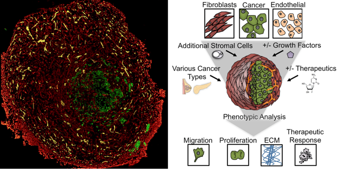
Bioprinted in vitro tumor models based on human cancer cells can accurately reproduce the characteristics of human cancer tissue (Image: Oregon Health & Science University).
Related Articles