


By Brinter Ltd.
Brinter®3D bioprinting technologies for cartilage repair
Brinter® strives to build technologies that advance diverse bioprinting applications for engineering and regenerating human tissues in osteoarthritis treatment and cartilage repair in knee, ankle, elbow and other injuries.
As one of the leading 3D bioprinting companies, Brinter® has been a proud partner in the EU-funded RESTORE project that has developed different bioprinting technologies and bio print heads, such as Rotary Tool and Multifluidics Tool, specifically for bioprinted cartilage patches for osteoarthritic knee treatment.
Bioprinting enables the precise patterning and assembling of cells and extracellular matrix (ECM) into functional 3D tissue constructs built from a patient’s own cells.
This technology will be a game changer in medicine, with bioprinting applications that range from drug screening and toxicology research to tissue and organ transplantation.
Basic Research In 3D printing tissue engineering
Bioprinting allows researchers to fabricate simplified homocellular tissue models for basic research or to produce more complex scaffolds with controlled spatial heterogeneity of physical properties, cellular composition, and ECM/biomolecule organization.
Developing novel 3D printing biomaterials and bioinks for 3D printing that are easily dispensable, mechanically robust, and able to maintain cellular viability are the keys to creating in vivo bioprinting technologies for direct patient treatment.
Bioprinted Tissues and Organs
A wide variety of tissues, such as blood vessels and neovascularized cartilage, have now been successfully bioprinted and the vision is to make it possible to 3D bioprint entire functional organs for those in need of organ transplant.
Several 3D bioprinting technologies have now been developed for cartilage repair, with the most useful techniques including:
- Inkjet bioprinting: using an inkjet bioprinter to precisely deposit cells and 3D printed biomaterials in a specific pattern to create an actual cartilage structure. This method allows for a high degree of control over the structure and organization of the printed tissue.
- Extrusion bioprinting: This technology uses pneumatic pressure to extrude a mixture of cells and biomaterials through a small nozzle to create the 3D structure of cartilage. This method is often used to create thicker and more complex structures.
- Laser-assisted bioprinting: Laser is either used to photopolymerize liquid photocurable bioink in a vat or deposit biomaterials onto a substrate and thus precisely shape 3D printing biomaterials into the desired 3D cartilage structure. This allows for a high degree of control over the structure and organization of the printed tissue, and can produce cartilage with high mechanical properties.
- Microscale bioprinting: 3D microfabrication techniques can create microscale 3D cartilage structures with high degree of control over the structure and organization of even the tiniest printed tissues, again with high mechanical properties.
- Scaffold-based bioprinting: A pre-made scaffold can be used as support for bioprinted cells to grow and differentiate into cartilage. This 3D bioprinting technology can use the patient’s own cells to fabricate cartilage with high mechanical properties but with minimal chances of rejection, with huge consequent savings on immunosuppressive drugs and their detrimental side effects.
All these technologies are still in the development stage and more research is needed to improve their efficiency and effectiveness. However, they have the potential to revolutionize the field of cartilage repair by providing a way to create replacement cartilage tissue that is biologically and mechanically similar to native cartilage.
Bioprinting in OA and cartilage repair
Even more advanced bioprinting solutions now on the horizon for cartilage repair and osteoarthritis (OA) therapy are reviewed in the soon-to-be-published Handbook of Surgical Planning and 3D Printing: Applications, Integration, and New Directions for which Brinter® CEO Tomi Kalpio and Bioprinting Product Manager Sanna Turunen were co-authors of the chapter ‘Future Solutions for Osteoarthritis using 3D Bioprinting of Articular Cartilage’.
The chapter explains how bioprinting offers an escape from the current dilemma in which the only treatment option remaining after immunosuppressive medication ceases to help people suffering from osteoarthritis (OA) is total knee replacement surgery with a prosthetic usually made of metal.
3D Bioprinting offers the prospect of creating customized cartilage patches with therapeutic properties to fill OA lesions. The chapter summarizes current scientific knowledge about 3D bioprinting cartilage and presents traditional and more advanced techniques, including novel biomaterials (bio inks) developed in the RESTORE Project that can be 3D printed to enhance the bioprintability of human cartilage to alleviate and combat OA.
‘It is reasonable to think that these designer nanoenabled bioinks with disease-modifying and regenerative properties could be used in the bioprinting of future medical products,’ the authors say, noting that while the vision of being able to bio print entire customized human organs from scratch remains out of reach, creating simpler tissues, such as blood vessels or cartilage, has already become a reality.
Principles of 3D Bioprinting
3D bioprinting is an additive manufacturing technique that uses a combination of living cells and biocompatible materials for “bio inks”, also known as bioinks, for layer-by-layer 3D printing of living structures.
The bioprinting process starts with the creation of a digital model, often based on Computer Tomography (CT) or Magnetic Resonance Imaging (MRI) scans or designed from scratch in CAD. The next step involves choosing relevant cell types and encapsulating 3D printing biomaterials. Selection of the bioink material is crucial, as it should mimic the complexity of the native ECM while having suitable physicochemical properties for the bioprinting process, i.e., possess good printability.
Traditional bioprinting techniques
The most frequently used technologies for 3D bioprinting include nozzle-based and laser/light-based techniques. Inkjet bioprinting is arguably the most common nozzle-based modality, while extrusion-based bioprinting is currently the most commonly used type of 3D bioprinting technology for tissue engineering applications. Unfortunately, single-material printing technologies are often unable to mimic the compositions, shape and architecture of native cartilage tissue accurately enough. Hence, more advanced variants of the standard bioprinting techniques have been developed.
The layer-by-layer 3D printing deposition of bioink is a particularly promising technique for engineering an organ or tissue. Mixing in cells either as single cells or as aggregates within the bioink represents a technological leap on the same scale as the original cloning from one somatic cell nucleus that produced Dolly the Sheep in 1997.
3D bioprints composed of human adipose stromal cells and hydrogel have been tested in rabbit knees and shown to regenerate osteochondral tissue. This chemistry-based hydrogel allowed for layer printing of different types of ECM hydrogels to form one multilayer construct without the need for chemical reagents or physical stimuli for crosslinking (Shim et al., 2016).
For cartilage injuries, 3D bioprinting of gelatin methacrylate, hyaluronic acid methacrylate, and human adipose-derived mesenchymal stem cells (hADSCs) resulted in human-like cartilage formation, with printing done with a Biopen handheld bioink 3D printing device that allows the simultaneous coaxial extrusion of bioscaffold and cultured cells directly into the cartilage defect in vivo in a single-session surgery.
Bioprinted hybrid constructs containing chondrocytes have also been tested for cartilage 3D printing tissue engineering applications in vivo in mice.
Chondrocytes, progenitor cells, stem cells, and even genetically modified cells have the most significant potential to repair damaged cartilage if bioprinted into 3D printed tissue engineering structures. However, cells from different sources have different properties and might not be suitable for reconstructing all types of cartilage tissues.
Future Techniques for Cartilage Bioprinting
Printed gradients of different cell types, biomolecules, and possible nanoparticles are often needed to properly mimic the characteristics of native tissues. Gradient printing requires precise control of shape, flow, composition and mixing of bioinks during the 3D bioprinting process. Therefore, new bioprinting techniques are constantly being researched and developed to overcome the challenges related to the printing of multicomponent bioinks into reasonably sized natural-like tissue structures with complex architecture. These more advanced approaches include:
- Coaxial / Microfluidic Bioprinting: The coaxial bioprinting (CBP) technique is a modified version of the EBB, and it has gained a lot of popularity due to its versatility and capacity to process a wide range of bioinks, even those with low viscosity. The concentrical nozzle allows the layer by layer 3d printing of multilayered or core-shell constructs from bioinks with rapid crosslinking mechanisms, e.g., those containing alginate. By developing custom slicing algorithms for 3D printing layer by layer, even inter-layer and intra-layer material switching and creating complex patterns within layers and across the entire construct is possible.
- Bioprinting in Support Bath: In this approach, mechanically unstable bioink is printed inside a gel-like consistency bath, which prevents the printed structure from collapsing. The bath can either be a crosslinking bath containing a crosslinker to trigger the gelation of the extruded bioink or supportive and act as temporary support for the ink. The technique is called FRESH bioprinting (Freeform Reversible Embedding of Suspended Hydrogels). This deposits the bioink as fibers into a gelatin support bath or slurry that is subsequently dissolved away after the bioprinting process. FRESH provides a strategy for fabricating non-self-supporting structures from water-rich, low-viscosity bioinks that could not otherwise be printed into 3D constructs.
- Volumetric Bioprinting: In contrast to all other bioprinting techniques that build objects through the sequential addition of building blocks, VBP-based technologies fabricate entire constructs at once, with clinically relevant arbitrary size and geometry within a time frame of a few seconds. The technique relies on the projection of a series of 2D patterned optical light fields within a volume of a photopolymer. These 2D light patterns act cumulatively to produce an optical 3D dose distribution triggering the polymerization of irradiated material into the desired object, demonstrating an ability to bio print features down to 80 µm using technical polymers, such as acrylates and elastomeric resin. However, VBP’s capabilities for 3D printing tissue engineering remain veiled as more studies need to be conducted to develop volumetric technologies for cell printing.
- In Situ Bioprinting: Also termed in vivo bioprinting, this refers to the direct deposition of bioinks at an injured site during a surgical operation to restore the living tissues. The technique makes the generation of scaffolds requiring in vitro preconditioning redundant as the body itself is used as a bioreactor. While obviously exciting as a surgical technique, it requires the development of highly advanced robotic bioprinters coupled with computer-aided design technologies (with scanners and interactive user interface for surgeons) as well as real-time monitoring systems and sensors for inspecting critical parameters, along with development of printed bioink that instantly solidifies in a living body without the use of external crosslinking agents such as ultraviolet light or chemical crosslinkers.
Future vision
The authors identify seven key takeaways from their studies:
- Articular cartilage (AC) cannot self-heal after being damaged due to osteoarthritis or traumatic injury. Without appropriate and well-timed intervention, the chondral defect may extend deep into the subchondral bone and require a joint replacement at the end stage.
- No effective drugs or treatment for damaged cartilage yet exist, requiring patients with AC damage to be treated with arthroplasty, involving widespread failure and complications, or undergo autologous chondrocyte implantation or microfracture surgery.
- As the functional regenerative repair of cartilage injuries remains a scientific and medical unmet need, significant steps have been taken to harness the possibilities of 3D bioprinting to produce cartilage patches.
- The design of the bioprinted cartilage substitute starts by defining the most suitable materials and the most appropriate bioprinting techniques to get the constructs of the desired shape, precision and functionality. Many materials, from soft natural hydrogels, such as alginate and collagen, to rigid synthetic polymers, such as polycaprolactone, have been used as bioinks in cartilage bioprinting.
- Layer-by-layer 3D printing can be used to construct a three-dimensional structure, starting from a 3D digital model. The more sophisticated bioprinting techniques allow the simultaneous deposition of multiple 3D printing biomaterials together with living cells and active biomolecules and enable the printing of complex and graded structures as well as the fabrication of personalized structures based on patient CT or MRI imaging.
- The selection of cells plays a vital role in facilitating the regenerative process of the printed tissue. Hence, cells capable of efficient proliferation as well as phenotype stability are the best choice for bioprinting. The most used cell types for cartilage regeneration are chondrocytes, mesenchymal stem cells (MSCs) and pluripotent stem cells (PSCs), which have the most significant potential to repair damaged cartilage.
- The heterogeneous and anisotropic tissue nature of cartilage has set challenges for reproducing its architecture in vitro by conventional bioprinting techniques. Therefore, in the last decades, researchers have moved towards more advanced bioprinting technologies to fabricate alternative functional cartilage constructs by introducing pioneering bioprinting techniques, including coaxial/microfluidic bioprinting, bioprinting in support bath, volumetric bioprinting, and in situ
They conclude: “The fast development of 3D bioprinting techniques gives hope to future therapies for cartilage injuries and OA, for which a clear need exists for more efficient treatment options. The rapid progress of 3D bioprinting platforms and novel options could revolutionize transplantation options for OA patients.
“In addition, advanced bioinks with improved printability and cellular compatibility are under development and could help halt disease progression. We foresee a 3D bioprinting revolution, and the development of future treatments is still in its infancy.”
Resources
Click on User-centered smart nanobiomaterial-based 3D matrices for chondral repair for further detail on the EU-funded RESTORE project.
Click on Brinter® Rotary Tool for further detail on technology for biological applications.
Click on Brinter® Multifluidics Tool for further detail on technology for biological applications.
Click on BioPrinting Applications for further information.
Click on Handbook of Surgical planning and 3D printing: applications, integration, and new directions for further details.
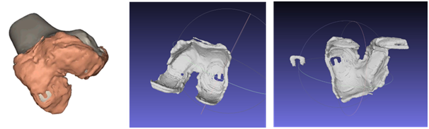
A representative 3D reconstructed model of the osteoarthritic knee joint from the RESTORE Project’s online database, together with STL models of the femoral cartilage with a horse-shoe-shaped cartilage patch designed to fit the lesion.
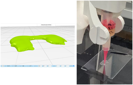
(Left): The horse-shoe-shaped cartilage patch model was created using various imaging techniques to unravel the 3D architecture of the tissues and control the positioning of print heads to place bioink in a 3D shape for a patient-specific match. (Right): 3D bioprinting process of the patch using Brinter® Rotary Tool print head and bioink material mixed with cells, spheroids or organoids.
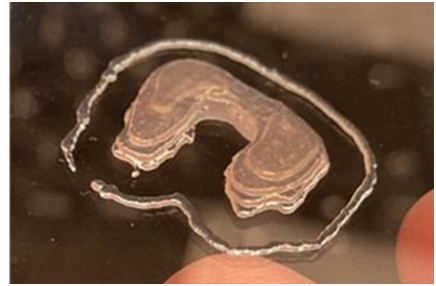
The completed cartilage patch bioprinted using the Brinter® Rotary Tool print head.
Related Articles