


By NanoMEGAS SPRL

Advanced Structural Characterization of Pharmaceutical Polymorphs using Micro Electron Diffraction
Brussels-based instrumentation hardware and software manufacturer NanoMEGAS (NM) is a specialist in advanced electron diffraction technologies in Transmission Electron Microscopy (TEM), including several patented technologies for phase/orientation mapping, strain mapping and 3D shape characterization of nanoparticles in liquids.
NM tools allow application of 3D Electron Diffraction (3D-ED)/ Micro Electron Diffraction (Micro-ED) for advanced characterization of pharmaceutical compounds.
Its proprietary technology platforms include the novel DigiSTAR digital precession unit that adds 3D Electron Diffraction to TEM units to enable them to characterize crystalline materials and solve nanocrystalline structures that are difficult to determine using conventional X-Ray diffraction techniques.
Micro-ED advantages
In recent years, the scientific community has shown a renewed interest in use of Micro-ED for characterizing those active pharmaceutical ingredients (APIs) for which it is challenging to grow suitable size crystals for single crystal X-ray diffraction. Traditional diffraction based techniques e.g. single-crystal x-ray diffraction (SCXRD) and Powder X-ray Diffraction (PXRD) have their own challenges, including:
- low crystallinity of the sample, which produces a broadening of peak profiles
- long cell parameters and pseudo symmetries, which lead to peak overlap even at low and medium resolution in PXRD
- presence of minor impurities or polymorphic forms.
In all these cases, 3D-ED/Micro-ED in TEM provides a useful alternative for structural studies, allowing crystals as small as 50 nm to be studied. The principle of acquiring 3D-ED data consists on focusing the electron beam on a micronic crystal in TEM/STEM mode and sampling the reciprocal space in small steps using beam precession or continuous rotation of the crystal.
NanoMEGAS recently presented a poster on application of these techniques to the 11th Conference on Crystal Forms in Bologna, Italy (CF@Bo11), authored by its scientists Partha Pratim Das, Athanassios S. Galanis, Alejandro Gómez Pérez, and Stavros Nicolopoulos.
Four Case Studies
The NanoMEGAS study highlights four recent case studies of investigations of pharmaceutical drugs using experimental 3D-ED that provided valuable new insights into the actual structures of these compounds, in some cases where the structure was not previously reported in literature.
These included:
- Carbamazepine Structure: Carbamazepine (CBZ) is primarily used in the treatment of epilepsy and neuropathic pain. It may be used in schizophrenia along with other medications and as a second-line agent in bipolar disorder. CBZ exists in several polymorphic forms. 3D-ED analysis of the carbamazepine compound provided an ab-initio solved molecular structure for the compound (Figure 1).
- Ramelteon Structure (in Colloboration with TeraCrystal): Ramelteon is the first selective melatonin MT1 and MT2 receptor agonist approved by the U.S. Food Drug and Administration (FDA) in 2005 for the treatment of insomnia. Again, 3D-ED analysis of the Ramelteon crystals provided solved molecular structure for the compound (Figure 2).
- Loratadine Form II Structure (in collaboration with Novartis) Loratadine, available under the brand name Claritin among others, is a medication used to treat allergies that exists in two different polymorphic forms. Ramelteon crystals Experimental 3D-ED TEM produced solved molecular structure (Figure 3).
- Linagliptin Structure (in Collobration with ICDD): Linagliptin, sold under the brand name Tradjenta inter allia , is a medication used to treat diabetes mellitus type2. At least 30 polymorphs of Linagliptin are known to exist. The 3D-ED data obtained by NanoMEGAS produced an ab-initio solved structure and closely matches Synchrotron data for the patented form for which no structure was previously reported (Figure 4).
The NM studies showed that 3D-ED/Micro-ED techniques in combination with Direct Detection cameras could be used as a powerful tool for phase identification and structural characterization for nm size (50-500nm) beam sensitive pharmaceutical materials.
The acquired 3D-ED data can be processed to determine ab-initio unit cell, space group, atomic positions and, moreover, hydrogen atom positions can also be determined.
Summary 3D-ED & Micro-ED benefits
In summary, NanoMEGAS 3D-ED/Micro-ED solutions allow a range of advanced applications that include:
- Full characterization of small molecule organics or impurities that cannot be studied by X-ray diffraction
- Polymorphic studies of organic pharmeceuticals
- Determination of unit cell, symmetry and crystal structure of nano crystals
- Determination of absolute configuration/Chrirality and hydrogen atom positions in organic pharmaceuticals
- Study of crystals in hydrated or solvated forms
Resources
Click on Electron Diffraction Tomography Micro-ED for Structural Characterization of Pharmaceutical Compounds to view original study in a poster.
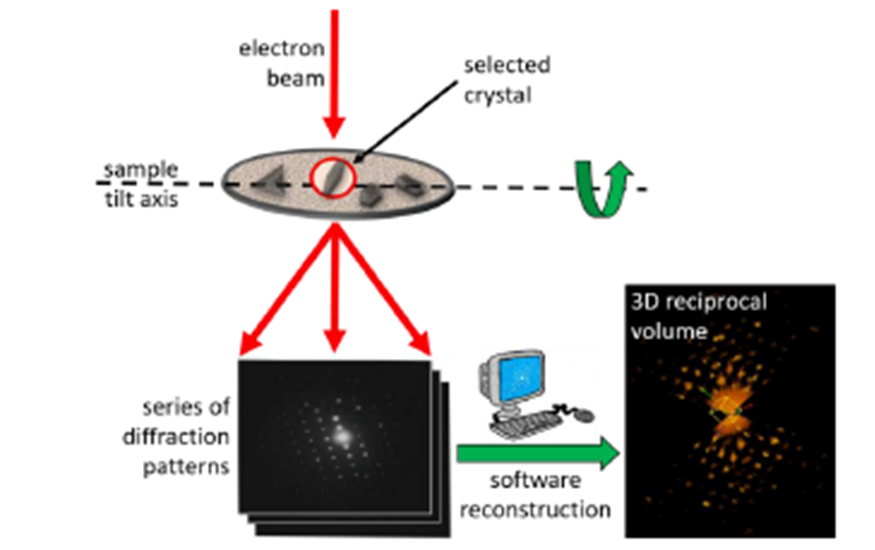
Principle of 3D ED data collection in TEM.
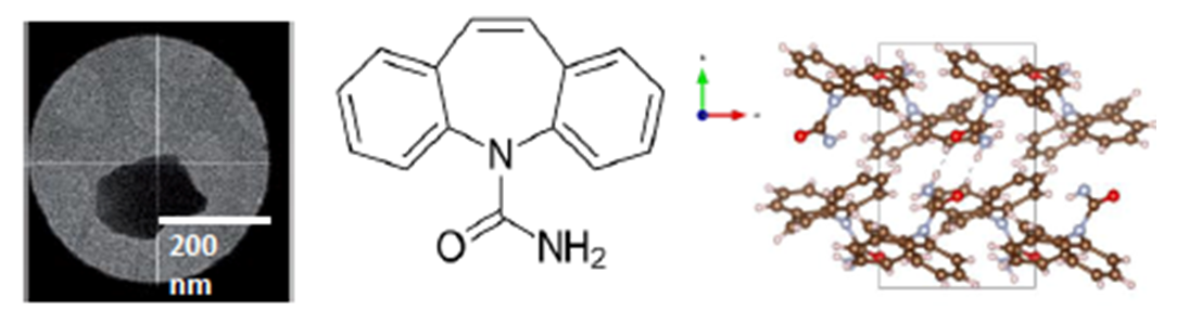
Figure 1: TEM Image of the carbamazepine nanocrystal and ab-initio solved structure from 3D-ED data at RT.
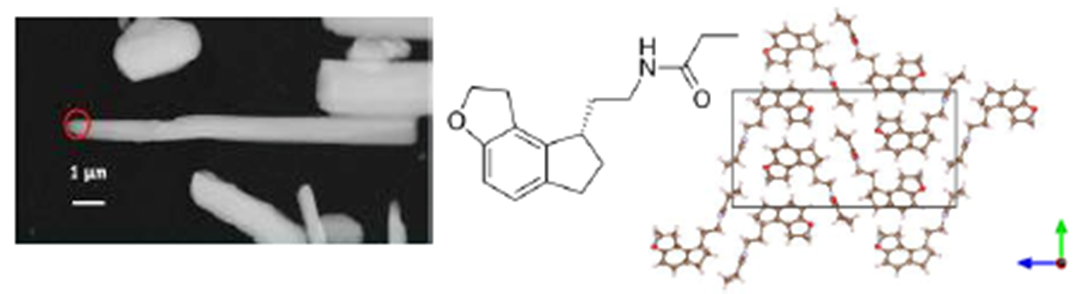
Figure 2. TEM Image of the Ramelteon crystals and solved structure from 3D-ED data at RT by Simulated Annealing.
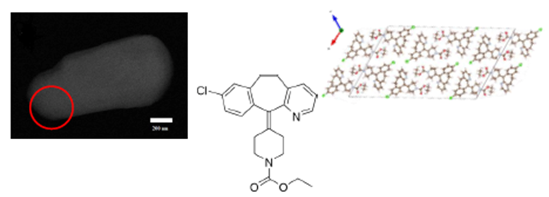
Figure 3. TEM Image of the metastable Loratadine Form II crystal and solved structure from 3D-ED data at RT by Simulated Annealing.
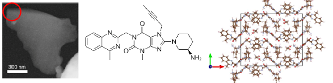
Figure 4. TEM Image of the Linagliptin crystal and ab-initio solved structure from 3D-ED data at RT.
Related Articles