


By NanoMEGAS SPRL

NanoMEGAS collaborates with Novartis Pharma on study using electron diffraction to solve metastable Loratadine crystal structure
Brussels, Belgium: – NanoMEGAS SPRL, the world-leading instrumentation hardware and software specialist and innovator in Transmission Electron Microscopy (TEM), has contributed its 3D Electron Diffraction (3D ED) technology to a study that sheds structural insights into the crystalline structure of loratadine as an example of studying metastable pharmaceutical nanocrystalline organic polymorphs.
Metastable polymorphs that typically display higher solubility than their thermodynamically stable counterparts, whilst having dissimilar mechanical and biopharmaceutical properties, have become a particular focus in drug development.
Multinational team
The study ‘Structural analysis of metastable pharmaceutical loratadine form II, by 3D electron diffraction and DFT+D energy minimization’ led by Dr. Grahame Woollam of Novartis Pharma AG, Basel, has been published in the Royal Chemistry Society’s journal CrystEngComm.[1]
NanoMEGAS researchers were part of the study team that also involved researchers from the Center for Nanotechnology Innovation (NEST) in Pisa, Italy, and Avant-garde Materials Simulation, based in Germany.
Maintaining polymorph stability and characterization method
The 3D ED study at room temperature succeeded to establish that the loratadine crystallites were pure phase with no other polymorphic form present, meaning that 3D data collected at room temperature had circumvented potential phase changes, thus preserving the metastable polymorph during structural elucidation.
The study establishes a promising protocol enabling the exploitation of combination of density functional theory and electron diffraction data with limited resolution obtained from beam sensitive organic materials that can be widely extended to a number of pharmaceutical compounds that are not amenable for the growth of large single crystals required by single crystal X-ray diffraction, and for which efficient structure determination is required.
3D ED data of three individual nanocrystals of loratadine form II were acquired at room temperature using Direct Detection Timepix (ASI, Netherlands) detector in STEM mode at very low illumination conditions with electron beam precession (Digistar hardware from NanoMEGAS SPRL , where ED intensities were processed using eADT software.
Overcoming TEM 3D ED limitations for limited resolution data
3D ED provides a robust means to determine the crystal structure of various nanocrystalline materials ranging from metals, alloys, minerals, , etc., to organic pharmaceuticals and proteins. Despite remarkable progress in 3D ED methods over recent years, the technique has not been applied specifically for structural elucidation of metastable forms of pharmaceutical materials, largely because the quantity and quality of acquired data on pharmaceutical compounds has been limited by the beam sensitivity of the materials small crystal size and possible occurrence of mosaicity and defects.
The study team say the significance of their work is to demonstrate how is possible to determine structure of a complex /metastable pharmaceutical compounds (even at room temperature) like loratadine starting from limited resolution 3D ED data, not suitable for performing an ab initio structure solution.
“We prove that in such cases the crystal structure of the pharmaceutical compound can be recovered by employing real space methods, like simulated annealing, where information about molecular connectivity and geometry of the rigid bodies is supplied a priori,” the study reports.
Extending the drug development database
The study concludes: “Our results reveal a very promising protocol enabling the exploitation of ED data with limited resolution obtained from beam-sensitive organic materials at room temperature. Our method uses very small quantity of material (eg mgr, much less quantity than X-Ray powder studies) and is widely extendable to a number of pharmaceutical compounds that cannot be readily crystallized and for which an efficient structure determination is required.”
This opens the way for many more polymorphs to be added to the CSD Cambridge Structural Database that forms the basis for ‘health checks’ to inform scientists whether a particular form is suitable for development.
“The addition of many more kinetically stable products captured within this nanoscale range where polymorphs with smooth surfaces may in fact be more stable will facilitate studies of crystallization and material science”.
“Access to structural information from kinetically driven products may help to rationalize molecular through to structural assembly by observing less common H-bonding networks, packing motifs or structural rugosity, illuminating stepping-stones on the crystallization pathway,” the study concludes.
Reference
- Woollam, G.R., Das, P.P., Mugnaioli, E., Andrusenko, I., Galanis, A.S., Streek, J. van de, Nicolopoulos, S., Gemmi, M. and Wagner, T. (2020). Structural analysis of metastable pharmaceutical loratadine form II, by 3D electron diffraction and DFT+D energy minimisation. CrystEngComm, [online] 22(43), pp.7490–7499. doi:10.1039/D0CE01216E.
About NanoMEGAS
Brussels-based instrumentation hardware and software manufacturer NanoMEGAS SPRL (NM) is a specialist and innovator in advanced electron diffraction technologies in Transmission Electron Microscopy (TEM), with exciting applications for the pharmaceutical industry.
NanoMEGAS produces and distributes hardware and software solutions for TEM applications that can be used for academic and industrial characterization of nanocrystalline and amorphous nanomaterials for pharmaceuticals and for polymorphic analysis. NM also provides scientific consulting for pharmaceutical companies wishing to characterize nano-crystalline and amorphous pharmaceuticals.
NanoMEGAS was founded in 2004 by a team of scientists and experts in the field of electron crystallography and catalysis. Inspired by pioneering work carried out on electron crystallography using precession electron diffraction at Bristol, UK at 1996 the NanoMEGAS team was first to develop and market its “spinning star” universal precession device for TEM in 2004. Today, there are more than 220 installations worldwide and more than 900 scientific publications related with NanoMEGAS applications & instrumentation in various fields, including pharmaceuticals.
Learn more at https://nanomegas.com
Resources
Click on Structural analysis of metastable pharmaceutical loratadine form II, by 3D electron diffraction and DFT+D energy minimization to access full study.
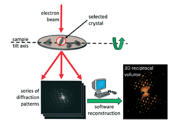
Sketch of the 3D ED method. One single nanocrystal is illuminated by a parallel electron beam while rotating continuously or in fixed steps around the goniometer axis of the TEM. The whole tilt range is used. Sequential non-oriented electron diffraction patterns are recorded and later reconstructed in a 3D volume by software. Finally, cell parameters, orientation matrix and reflection intensity integration are performed.
Related Articles