


By NanoMEGAS SPRL

Novel Pharmaceutical Applications using Electron Diffraction
NanoMEGAS (NM) is a specialist in advanced precession electron diffraction (PED) technologies in Transmission Electron Microscopy (TEM).
Precession electron diffraction (PED) is a specialized method to collect TEM diffraction patterns in a transmission electron microscope. By rotating (precessing) a tilted incident electron beam around the central optical axis of the microscope, a PED pattern is formed by integration over a collection of diffraction conditions. This produces a quasi-kinematical electron diffraction pattern which is more suitable as input into direct methods algorithms to determine the crystal structure of the sample.
PED theory
Precession electron diffraction can utilize standard TEM instrument configuration, using the beam tilt coils located pre-specimen to move the electron beam off of the optic axis so it is incident with the specimen at an angle.
The resulting electron diffraction pattern consists of a summation or integration over the patterns generated during precession. While the geometry of this pattern matches the pattern associated with a normally incident beam, the intensities of the various reflections approximate those of the kinematical pattern much more closely.
History of PED
The first precession electron diffraction system was developed in Bristol, UK, in the early 1990s, with feasibility of the technique demonstrated by preliminary investigation into the Er2Ge2O7 crystal structure by reducing dynamical effects and providing quasi-kinematical patterns which helped to solve the structure using direct methods. Over the next decade, a number of university groups in Norway, Italy and the USA developed their own precession systems and verified the technique by solving more complex crystal structures.
In 2004, NanoMEGAS developed the first commercial precession system capable of being retrofit to many modern TEM. This hardware solution enabled more widespread implementation of the technique and spurred its more mainstream adoption into crystallography. As of 2023, more than 900 published studies have relied on the technique to investigate nanocrystalline materials most of them could not be studied by other conventional crystallography techniques like X-Ray diffraction. NanoMEGAS precession systems is used by more than 240 laboratories across the world.
PED Advantages
PED techniques is advantageous in investigating crystal structures. These include:
- Quasi-kinematical electron diffraction patterns:While the underlying physics of the electron diffraction (ED) is still dynamical in nature, the conditions used to collect PED patterns minimize many of these effects. The scan/de-scan procedure reduces ion channeling because the pattern is generated off of the zone axis. Integration via precession of the beam minimizes the effect of non-systematic inelastic scattering, such as Kikuchi lines. Few reflections are strongly excited at any moment during precession, and those that are excited are generally much closer to a two-beam condition. Furthermore, for large precession angles, the radius of the excited Laue circle becomes quite large. These contributions combine such that the overall integrated electron diffraction pattern resembles the kinematical pattern much more closely than a single zone axis pattern.
- Broader range of measured reflections:The Laue circle (see Ewald sphere) that is excited at any given moment during precession extends further into reciprocal space. After integration over multiple precessions, many more reflections in the zeroeth order Laue zone (ZOLZ) are present, with many more close to kinematical intensities. This provides considerably more information to input into direct methods calculations, improving the accuracy of phase determination algorithms. The higher order Laue zone (HOLZ) reflections present in the pattern can provide more complete information about the three-dimensional nature of reciprocal space, even in a single two-dimensional PED pattern.
- Practical robustness:PED is less sensitive to small experimental variations than other electron diffraction techniques. Since the measurement is an average over many incident beam directions, the pattern is less sensitive to slight misorientation of the zone axis from the optic axis of the microscope, and resulting PED patterns will generally still display the zone axis symmetry. The patterns obtained are also less sensitive to the thickness of the sample, a parameter with strong influence in standard electron diffraction patterns.
- Very small probe size:Because X-Rays interact so weakly with matter, there is a minimum size limit (>> 1 micron) for single crystals that can be examined via x-ray diffraction methods. In contrast, electrons can be used to probe much smaller nano-crystals in a TEM. In PED, the probe size is limited by the lens aberrations and sample thickness. With typical TEM-FEG aberration corrected TEMs, the minimum probe size is usually around 0.9-1 nm.
Precession electron diffraction applications
Precession electron diffraction can be used for a range of different applications including orientation mapping, phase mapping, strain mapping, diffraction tomography and spectroscopy.
PED is typically conducted using accelerating voltages between 100-400 kV. Patterns can be formed under parallel or convergent beam electron diffraction (CBED) conditions. Most modern TEMs can achieve a tilt angle, φ, ranging from 0-3°. Precession frequencies can be varied from Hz to kHz, but in standard cases 100 Hz has been used .In choosing a precession rate, it is important to ensure that many revolutions of the beam occur over the relevant exposure time used to record the electron diffraction pattern. This ensures adequate averaging over the excitation error of each reflection. Beam sensitive samples may dictate shorter exposure times and thus, motivate the use of higher precession frequencies.
Resources
Click on Precession Electron Diffraction and Applications for further information.
Click on NanoMEGAS Bibliography to access more than 900 articles of Electron Beam Precession and Applications literature.
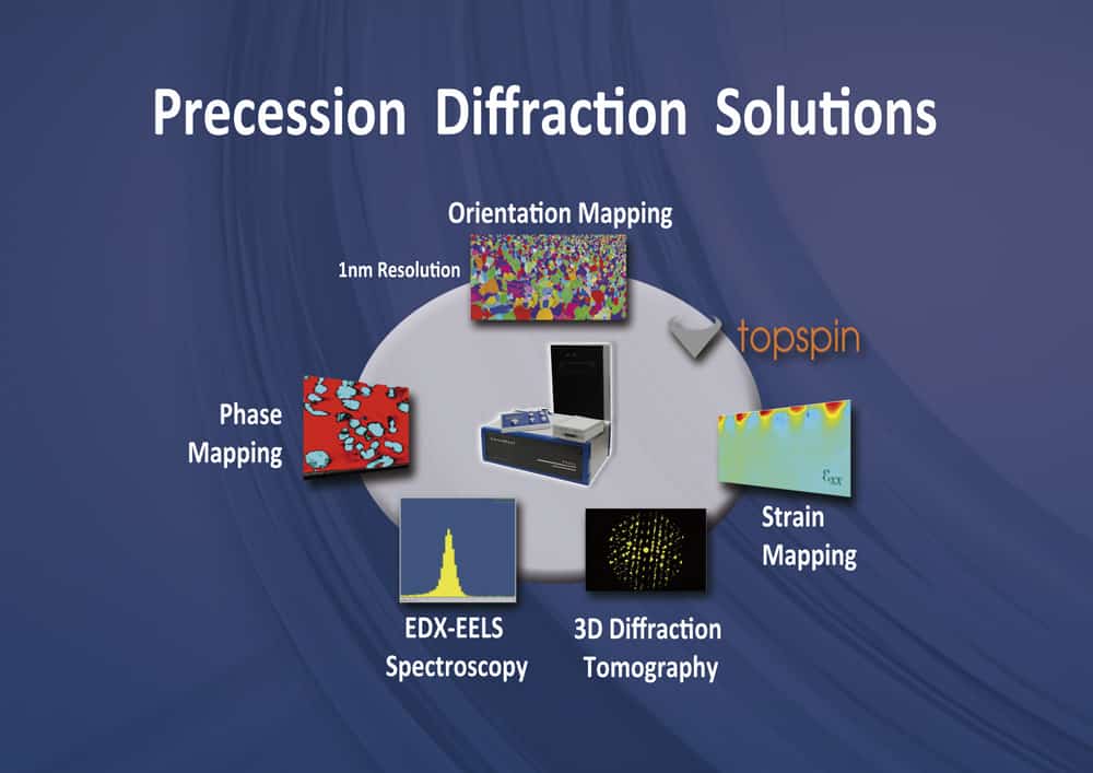
PED can be used for a range of different applications including phase and orientation mapping, diffraction tomography and spectroscopy.
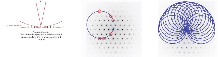
Precession diffraction geometry (left) and Laue circle sweeping through the reciprocal space due to PED movement (right).
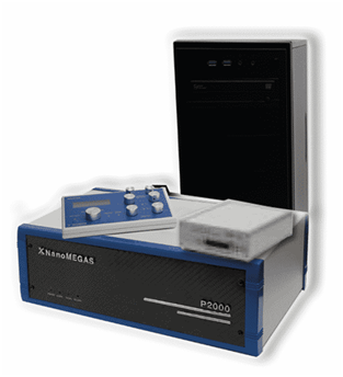
Digistar digital precession device with workstation (adaptable to most modern TEM , including single or double corrected Cs) and various modern TEMs where has been installed.
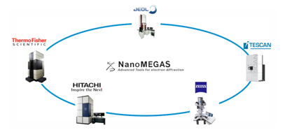
NanoMEGAS commercial procession system that can be fitted to all modern TEM microscopes.
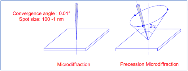
Precession electron diffraction basic theory: Rotating (precessing) a tilted incident electron beam around the optical axis of the TEM microscope forms a PED pattern by integrating a collection of diffraction conditions to produce a quasi-kinematical diffraction pattern.
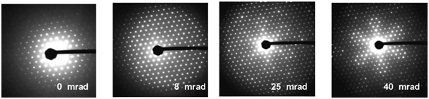
From left to right: [111] Electron diffraction pattern of mayenite mineral (cubic a= 12.5 A) at different precession angles 0 mrad for complete dynamical ED pattern, 40 mrad precession angle for quasi-kinematical electron diffraction (ED) pattern.
Related Articles