


By BIOSYNTH AG

AquaSpark™ beta-D-glucuronide stand-alone chemiluminescent detection of GUS and E. coli
Abstract: A superior probe for E. coli identification
Microbiology and organic fine biochemical CDMO and specialist supplier Biosynth AG, has developed an innovative dioxetane-based AquaSpark™ chemiluminescent β-glucuronidase probe that shows significant advantages over traditional chromogenic and fluorogenic detection methods for β-glucuronidase (GUS).
The AquaSpark™ beta-D-glucuronide (A-8175_P00) is the first single-compound chemiluminescent substrate for E. coli detection. Upon hydrolytic cleavage the AquaSpark™ probe spontaneously emits a green light that allows for the sensitive detection of β-glucuronidase activity that is a primary indicator of E. coli contamination.
Ultra-fast and responsive assays using AquaSpark™ β-glucuronidase substrates for E. coli detection allows for faster detection of bacterial contamination in food and healthcare sectors as well as in the environment.
Background: β-glucuronidase and E. coli
A member of the glycosyl hydrolase family, the β-glucuronidase enzyme (beta D-glucuronoside glucuronosohydrolase; EC 3.2.1.31) cleaves beta-D-glucopyranosuronic residues liberating a molecule of glucuronic acid and the remaining sugar molecule or aglycon.
The expression, sequence, structure, and function of human β-Glucuronidase (GUSB) have been comprehensively studied over the past decades, revealing that GUSB is an important lysosomal enzyme involved in the degradation of glucuronate-containing glycosaminoglycan. GUSB deficiency results in the accumulation of mucopolysaccharides in humans and is the leading cause for Mucopolysaccharidosis VII (‘Sly syndrome’).
In prokaryotes, the β-glucuronidase of Escherichia coli (GUS) is also well characterized. In the bacterial world it occurs almost exclusively in Escherichia coli (E. coli), the gram negative bacterium that inhabits the intestines of warm-blooded animals and is always found in feces.
The presence of E. coli is the most important indicator of fecal contamination of drinking water, surface water, food and beverages. Certain strains of E. coli are also human pathogens. Living cells of E. coli can be reliably detected with media that contain a substrate for β-glucuronidase, an enzyme that occurs in the microbial world mainly but with a few exceptions in E. coli. Many state-of-the-art E. coli tests are using this enzyme with defined substrates (Rice et. al, 1990).
The E. coli GUS protein is 603 amino acid residues long and shares about 50 percent sequence identity with the human β-glucuronidase with same substrate specificity.
The 4-Methylumbelliferyl beta-D-glucuronide (MUG) substrate is frequently used for detecting β-glucuronidase and is recommended in international guidelines as the fluorogenic substrate for E. coli detection.
GUS assays in plant molecular biology
The β-glucuronidase enzyme from E. coli (GUS) is one of the most popular reporter gene assays in plant molecular biology as it shows several useful characteristics:
- There is no detectable β-glucuronidase activity in higher plants that could otherwise produce background signals.
- The enzyme is stable and tolerates commonly used detergents and reducing agents (such as ME or DTT) in the assay procedures.
- The enzyme is also capable of tolerating amino-terminal additions, making it useful for the study of plant organelle transport.
The GUS gene encoding β-glucuronidase is frequently used to analyze the activity of a promoter either quantitatively or through visualization of its activity in different tissues. A wide range of plasmids that serve as plant GUS expression vectors is commercially available.
Biosynth’s chromogenic enzyme substrate X-gluc (5-bromo-4-chloro-3-indoxyl-beta-D-glucuronic acid (Biosynth Cat. No. B-7300) is traditionally applied in ‘GUS staining’ of genetically modified plants. The enzyme acts by cleaving the β-glucuronic acid from the substrate X-gluc and thus producing a blue color in transformed tissues. As X-gluc forms a blue precipitate it is mostly used for localization studies, while Biosynth’s fluorescent substrate MUG (4-Methylumbelliferyl-beta-D-glucuronic acid (Cat. No. M-5700) is typically used to measure the level of GUS gene expression.
E. coli detection using AquaSpark™ beta-D-glucuronide
Biosynth developed its strong emitting dioxetane-based AquaSpark™ chemiluminescence technology in partnership with Prof. Doron Shabat at the Tel Aviv University. The novel AquaSpark™ probes have significant advantages over conventional methods, bringing detection of specific enzyme activities to a level of sensitivity comparable to bioluminescent assays without the need of light producing enzymes (Luciferase).
This allows the AquaSpark™ beta-D-glucuronide probe (Biosynth Cat. No. A-8175_P00) to detect specific β-glucuronidase activity with unprecedented sensitivity, significantly out-performing the previous gold standard 4-Methylumbelliferyl-beta-D-glucuronic (MUG) acid.
Furthermore, the AquaSpark beta-D-glucuronide highly sensitive chemiluminescent substrate can be used to identify living E. coli after incubation periods as short as six hours.
In testing, β-glucuronidase positive E. coli and β-glucuronidase negative Klebsiella pneumonia cultures were inoculated in test tubes containing diluted Luria Bertani (LB) broth with an inducer for β-glucuronidase . After 6 hours of cultivation at 37°C , samples were drawn from the cultures and the bacteria were lysed for 30 minutes.
AquaSpark™ beta-D-glucuronide (50 µM) was added to the E. coli, the K. pneumonia lysate and to a sterile control solution. Chemiluminescence light emission was recorded for 20 minutes and the maximum emissions (relative light units, RLU) were used for quantification.
While E. coli samples produced a strong light signal β-glucuronidase negative Klebsiella pneumonia did not produce a chemiluminescence signal higher than the sterile control.
The time factor is highly significant since speed is a crucial factor in food and water control as well as in clinical analytics. AquaSpark™ chemiluminescence-based β-glucuronidase assays provide a new capability to detect E. coli bacteria contamination, even at low levels of E. coli -specific enzyme activity, much earlier in comparison to traditional bacteria stains or fluorescent dyes.
Reference:
Rice, E.W., Allen, M.J. and Edberg, S.C. (1990) Appl Environ Microbiol. 56 (5): 1203–1205. Efficacy of beta-glucuronidase assay for identification of Escherichia coli by the defined-substrate technology.
Resources
Click on Biosynth AquaSpark™ GUSB testing for E. coli for more information.
Click on BIOSYNTH to contact the company directly.
Click on AquaSpark™ beta-D-glucuronide, 10 mM in DMSO (A-8175_P00) to see product details.
Click on Product Information Sheet for more technical information.
Click on AquaSpark™ substrates for animated video.
Click on Biosynth newsletter to subscribe.
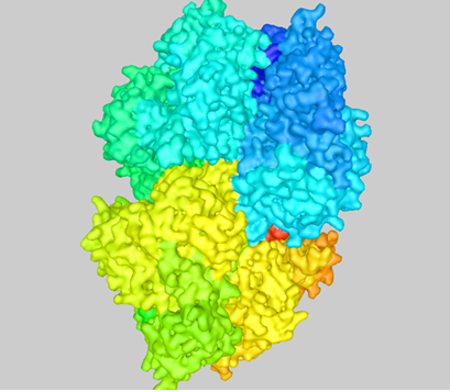
Structure of E. coli β-glucuronidase

Efficacy of beta-glucuronidase assay for identification of Escherichia coli by the defined-substrate technology
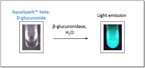
Luminescent β-glucuronidase tests using AquaSpark™
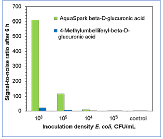
Comparison of signal-to-noise ratios achieved after 6 hours of E. coli cultivation, using either AquaSpark™ beta-D-glucuronide or MUG (4-Methylumbelliferyl-beta-D-glucuronic acid) for the detection of specific β-glucuronidase activity.
Supplier Information
Supplier: BIOSYNTH AG
Address: Rietlistr. 4, 9422 Staad, Switzerland
Tel: +41 (0)71 858 20 20
Fax: +41 (0)71 858 20 30
Website: www.biosynth.com
Related Articles